
Key Takeaways
- Tumors are classified by the WHO CNS5 classification, which combines how cells look under a microscope with their genetic makeup.
- Grades range from 1 to 4, with higher numbers indicating faster growth and higher malignancy.
- Molecular markers, like IDH mutations, now play a bigger role in prognosis than traditional imaging alone.
- Treatment is usually "multimodal," meaning a combination of surgery, radiation, and chemotherapy.
- New targeted therapies, such as vorasidenib, are improving outcomes for specific genetic subtypes.
The Modern Way We Classify Brain Tumors
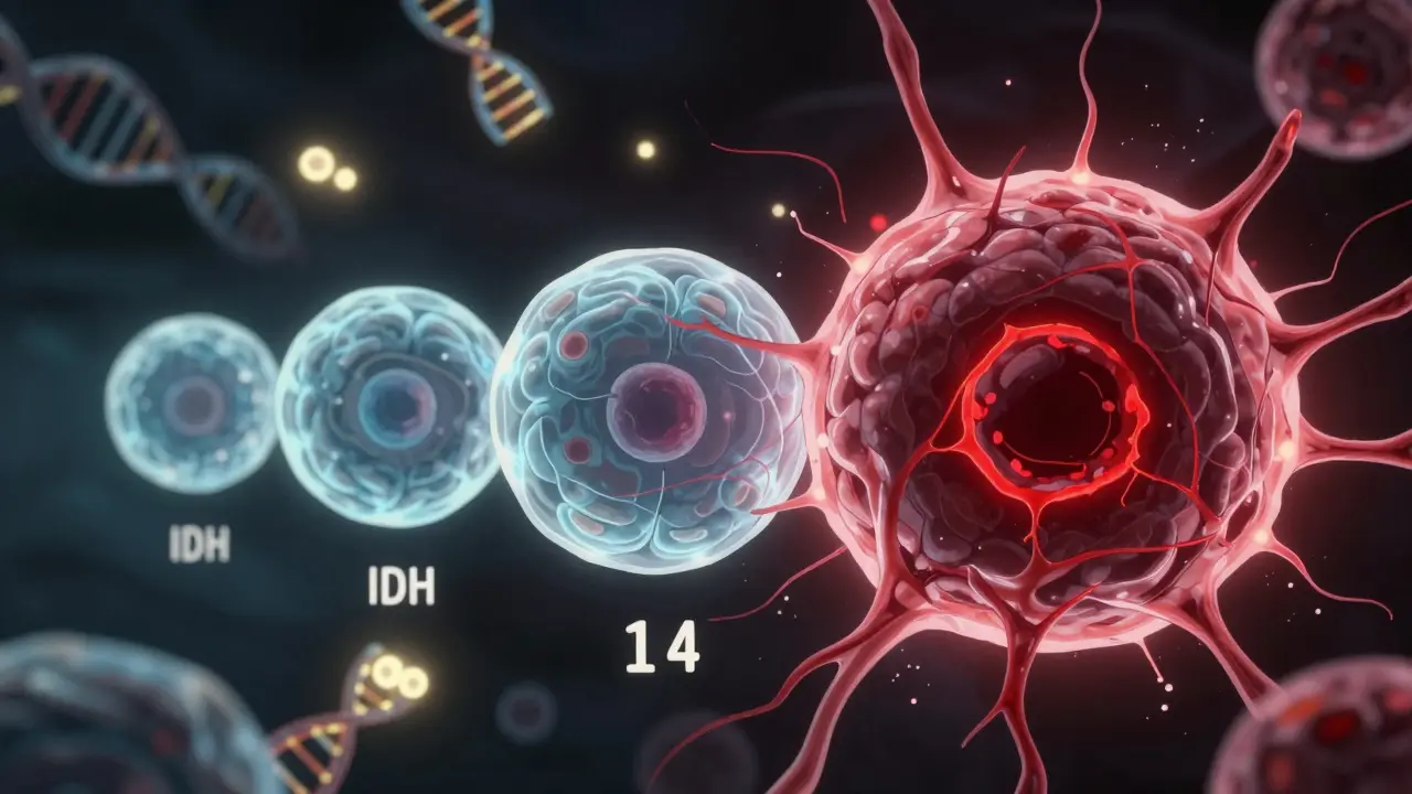
For a long time, doctors graded tumors simply by looking at them under a microscope. If the cells looked chaotic and grew fast, it was a high grade. But we've learned that two tumors looking identical under a lens can behave completely differently. That's why the WHO CNS5 classification is the current global standard for classifying tumors of the central nervous system. Released in 2021, this system shifted the focus toward molecular diagnostics.
Basically, the WHO CNS5 doesn't just ask "What does it look like?" but "What is its genetic signature?" For example, an Astrocytoma is no longer just a general category. Doctors now look for the IDH mutation. If a tumor is "IDH-mutant," it generally has a better prognosis than one that is "IDH-wildtype," even if they are both high-grade. This shift has improved diagnostic accuracy by as much as 40%, meaning patients get treatments tailored to their specific genetic markers rather than a one-size-fits-all approach.
Breaking Down the Grades: From 1 to 4
When you hear a "grade," think of it as the tumor's speed limit. A grade 1 tumor is like a slow walker; a grade 4 is like a race car. These grades help doctors predict how the tumor will behave and how likely it is to come back.
Grade 1 and 2 (Low Grade): These are typically slow-growing. Grade 1 tumors often have very distinct edges, making them easier for surgeons to remove entirely. Grade 2 tumors are a bit more sneakily integrated into the surrounding brain tissue. While they aren't as aggressive, they can sometimes "evolve" or transform into higher-grade tumors over time.
Grade 3 and 4 (High Grade): These are malignant. Grade 3 tumors, often called "anaplastic," show active proliferation-they are dividing and invading nearby healthy tissue rapidly. Grade 4 tumors are the most aggressive. They often create their own blood vessels to feed their growth and can develop necrotic (dead) centers because they grow so fast they outstrip their own blood supply. The most well-known grade 4 tumor is Glioblastoma, which accounts for over half of all malignant gliomas in adults.
| Grade | Growth Rate | Behavior | Common Example |
|---|---|---|---|
| 1 | Very Slow | Well-defined edges, low recurrence | Pilocytic Astrocytoma |
| 2 | Slow | Can infiltrate tissue, may upgrade | Low-grade Oligodendroglioma |
| 3 | Fast | Aggressive invasion of nearby tissue | Anaplastic Astrocytoma |
| 4 | Very Fast | Rapid multiplication, necrotic centers | Glioblastoma |
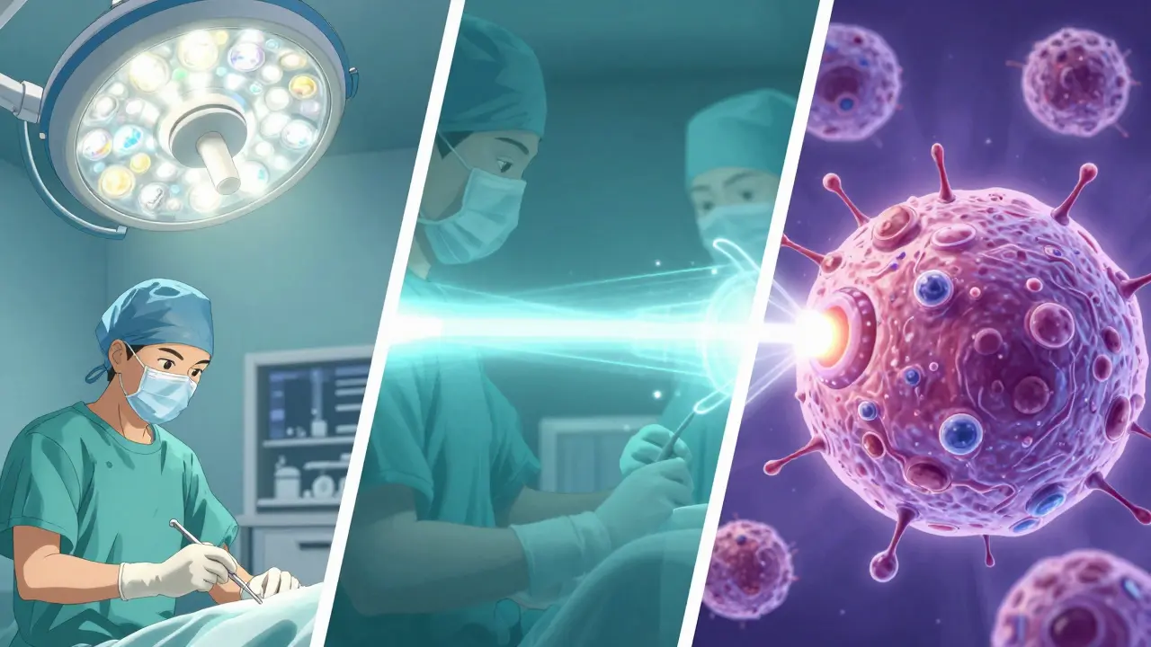
Multimodal Treatment: The Three-Pronged Attack
Because brain tumors are stubborn and often infiltrate healthy tissue, a single treatment rarely does the trick. Instead, doctors use multimodal therapy-a combination of different tools used in a specific sequence to maximize the chance of success.
- Surgical Resection: The first goal is usually "maximal safe resection." The surgeon removes as much of the tumor as possible without damaging critical parts of the brain (like speech or motor centers). In low-grade tumors, surgery can sometimes be a cure. In high-grade tumors, it's about reducing the tumor's mass to make the next treatments more effective.
- Radiation Therapy: High-energy beams are used to kill the remaining cancer cells that the surgeon couldn't see or reach. This is often focused precisely on the area where the tumor was located.
- Chemotherapy: Drugs like Temozolomide are frequently used. This is a systemic treatment that can cross the blood-brain barrier to attack cells that are still dividing. For glioblastoma, the "Stupp Protocol" (a mix of radiation and chemotherapy) has been the gold standard for years.
The Role of Molecular Markers in Treatment
The real breakthrough in the last few years is targeted therapy. We are moving away from "blunt" instruments like general chemo and toward "smart" drugs. For example, if a patient has an IDH-mutant grade 2 glioma, they might be eligible for a drug called vorasidenib. In the INDIGO clinical trial, this drug significantly extended the time patients lived without the tumor growing compared to those who didn't take it.
Another critical marker is MGMT promoter methylation. If the MGMT gene is "methylated" (essentially turned off), the tumor is often much more responsive to chemotherapy. This tiny piece of molecular data can completely change the outlook for a patient, shifting the conversation from "minimal hope" to "a strong chance of response." This is why molecular testing, though it can add a few thousand dollars to the bill and take a week or two to process, is non-negotiable in modern care.
Practical Realities and Patient Hurdles
Beyond the medicine, there is the human side of this experience. One of the hardest parts is the "diagnostic gap." Many patients wait weeks for a final pathology report because molecular testing is complex. It's not uncommon for a patient to have surgery on Monday but not know the exact grade and molecular subtype of their tumor for another ten days.
There's also a significant amount of confusion around the numbers. Some patients mistakenly believe that a "grade 2" means they have a "20% chance of survival," which isn't how grading works. Grade refers to the cellular speed and aggression, not a percentage of survival. Understanding this distinction is crucial for mental health and realistic planning. For younger patients, a diagnosis often brings up urgent life questions-such as fertility preservation-that must be addressed in the very narrow window before chemotherapy or radiation begins.
What's on the Horizon?
We are entering an era of "liquid biopsies." Instead of drilling into the skull for a tissue sample, researchers are finding ways to detect tumor DNA in the cerebrospinal fluid. A recent study showed nearly 89% sensitivity in detecting these markers, which could make monitoring for recurrence much less invasive.
Furthermore, the CODEL trial is currently looking at how combined chemotherapy can improve outcomes for those with oligodendrogliomas. The goal is to move toward a future where a brain tumor is treated more like a chronic disease-managed with targeted pills and precise monitoring-rather than an acute crisis managed with massive surgery and radiation.
What is the difference between a primary and secondary brain tumor?
A primary brain tumor starts in the brain tissue itself. A secondary (or metastatic) tumor starts elsewhere in the body-like the lungs or breast-and spreads to the brain. The grading systems discussed here, like the WHO CNS5, primarily apply to primary tumors.
Does a Grade 4 diagnosis mean the tumor cannot be treated?
No. While Grade 4 tumors are the most aggressive, they are treated with aggressive multimodal therapy. Some patients respond very well to the combination of surgery, radiation, and targeted drugs, and molecular markers (like IDH status) can significantly change the expected outcome.
How long does it take to get the final grade of a tumor?
Typically, it takes about 7 to 10 business days from the biopsy. This is because the tissue must be processed histologically and then undergo molecular testing to check for mutations like IDH, which takes extra time in the lab.
What is the 'Stupp Protocol'?
The Stupp Protocol is the standard of care for glioblastoma. It involves the surgical removal of as much of the tumor as possible, followed by a combination of radiotherapy and the chemotherapy drug temozolomide.
Can a low-grade tumor become high-grade?
Yes, this is known as "malignant transformation." Some Grade 2 tumors can evolve into Grade 3 or 4 tumors over time. This is why regular MRI monitoring is essential even for patients with slow-growing tumors.
Next Steps and Troubleshooting
If you or a family member has just received a diagnosis, the first step is to ensure you have the full molecular report, not just the surgical summary. Ask your neuro-oncologist specifically about IDH status and MGMT methylation, as these dictate which clinical trials or targeted drugs (like vorasidenib) you might qualify for.
If you are facing a long wait for a pathology report, don't panic. This delay is usually due to the precision required for molecular sequencing. In the meantime, focus on "quality of life" preparations and discuss fertility or long-term planning with your medical team before the first round of intensive treatment begins.


Stephen Luce
April 5, 2026 AT 23:09It's just so overwhelming to deal with these terms when you're already scared out of your mind. The part about the diagnostic gap really hits home because that waiting period for pathology is absolute torture for the family.
Victoria Gregory
April 6, 2026 AT 21:18Sending so much love and light to everyone reading this!!! 🌸✨ It's just wild how much a tiny genetic marker can change everything... truly makes you think about the mystery of life and how we're all just biological puzzles!!! 🧩💖🌈
Darius Prorok
April 8, 2026 AT 18:03I already knew about the Stupp Protocol. It's basic stuff.
Benjamin cusden
April 10, 2026 AT 11:43While the summary is adequate for a layperson, it glosses over the nuanced interplay between the blood-brain barrier and the pharmacological efficacy of temozolomide. One would hope the author understands that 'molecular markers' is a broad term and the specific epigenetic modifications are where the actual science resides.
dwight koyner
April 10, 2026 AT 22:51For those navigating this, please ensure you request a multidisciplinary team review. Having a neuro-surgeon, a neuro-oncologist, and a neuroradiologist discuss your case in a single room often leads to a more cohesive treatment plan than seeing them individually.
Ethan Davis
April 11, 2026 AT 13:27Sure, tell us about the 'new drugs' while the pharmaceutical companies keep the real cures hidden to make more money off the long-term chemo. These 'clinical trials' are just a way to test things on us without giving us the full picture of what's actually possible.
Kathleen Painter
April 11, 2026 AT 13:38I think it's really important for us to remember that while the science is evolving and the WHO CNS5 classification is a huge step forward, the emotional toll of this journey is something that a pathology report simply cannot capture, and we should all try to hold space for the people who are currently in that ten-day waiting period, because the anxiety of not knowing the grade of the tumor can be almost as paralyzing as the diagnosis itself, and maybe if we focus more on the community support side of things, the medical jargon won't feel quite so isolating for the newcomers in this struggle.
Jamar Taylor
April 12, 2026 AT 19:40Keep fighting! You've got this!
Ruth Swansburg
April 13, 2026 AT 11:09Stay strong. You are capable of overcoming this.
Windy Phillips
April 14, 2026 AT 23:43It is truly fascinating that some people actually believe these 'modern' treatments are a miracle cure... though I suppose the desperation of a Grade 4 diagnosis makes one susceptible to any glimmer of hope, no matter how statistically insignificant it may actually be!!!
Jitesh Mohun
April 15, 2026 AT 19:06stop crying and just get the molecular report done if you want to survive dont waste time on emotions just follow the data and get the surgery and the chemo and move on